Full Download Atlas of Clinical Imaging and Anatomy of the Equine Head - Larry Kimberlin | PDF
Related searches:
Atlas of Clinical Imaging and Anatomy of the Equine Head Wiley
Atlas of Clinical Imaging and Anatomy of the Equine Head - Kindle
Atlas of Clinical Imaging and Anatomy of the Equine Head on Apple
Atlas of Clinical Imaging and Anatomy of the Equine Head – Bóksalan
[PDF] Atlas Of Clinical Imaging And Anatomy Of The Equine
Atlas of Clinical Imaging and Anatomy of the Equine Head PDF
ATLAS - The University of Auckland - Medical and Health Sciences
Atlas of Clinical Imaging and Anatomy of the Equine Head by
Atlas of clinical imaging and anatomy of the equine head
Full Trial Atlas of Clinical Imaging and Anatomy of the
The Equine Distal Limb: An Atlas of Clinical Anatomy and
e-Anatomy: radiologic anatomy atlas of the human body - IMAIOS
Pocket Atlas of Sectional Anatomy: Computed Tomography and
Cross Sectional Anatomy By Ct And Intro To Procedures - NACFE
Trial New Releases Atlas of Clinical Imaging and Anatomy of
Imaging Anatomy of the Human Brain: A Comprehensive Atlas
Expanding the Computational Anatomy Gateway from clinical
e-Anatomy: The Interactive Atlas of Human Anatomy - University of
RadiologyEbooks.com: Free Radiology Apps and Radiology Ebooks
Anatomy, medical imaging and e-learning for healthcare
Abrahams’ and McMinn’s Clinical Atlas of Human Anatomy 8th
Ultimately the improved understanding you can acquire using this atlas can enhance clinical understanding and have a positive impact on patient care.
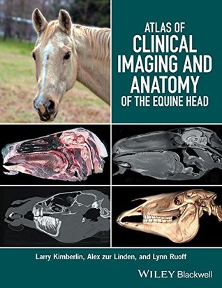
Atlas of clinical imaging and anatomy of the equine head presents a clear and complete view of the complex anatomy of the equine head using cross-sectional imaging. Provides a comprehensive comparative atlas to structures of the equine head pairs gross anatomy with radiographs, ct, and mri images presents an image-based reference for understanding anatomy and pathology covers radiography.
Medical image computing (mic) is an interdisciplinary field at the intersection of computer while closely related to the field of medical imaging, mic focuses on the atlas-based segmentation: for many applications, a clinical expe.
Atlas of upper and lower extremity muscles with origins and insertions. E- anatomy, interactive cross sectional imaging atlas of human.
Interactive anatomical atlas of the head, brain, and neck based on anatomical diagrams and ct and mri medical imaging exams.
Atlas of clinical imaging and anatomy of the equine head presents a clear and complete view of the complex anatomy of the equine head using cross-sectional imaging. � provides a comprehensive comparative atlas to structures of the equine head pairs gross anatomy with radiographs, ct, and mri images presents an image-based reference for understanding anatomy and pathology covers radiography.
This atlas achieves its goal to be a helpful pocket reference, rather than a textbook� there are few other such texts available for musculoskeletal imaging, with.
Atlas of clinical imaging and anatomy of the equine head presents a clear and complete view of the complex anatomy of the equine head using cross-sectional imaging. Provides a comprehensive comparative atlas to structures of the equine head; pairs gross anatomy with radiographs, ct, and mri images.
Anatomy in diagnostic imaging book description� now in its third edition, anatomy in diagnostic imaging is an unrivalled atlas of anatomy applied to diagnostic imaging. The book covers the entire human body and employs all the imaging modalities used in clinical practice; x-ray, ct, mr, pet, ultrasound and scintigraphy.
Vet-anatomy the interactive atlas of veterinary anatomy� vet-anatomy is a veterinary atlas of anatomy based on veterinary imaging (mri, ct, x-rays) and medical illustrations, designed and created by professional anatomists and veterinary imaging specialists.
Atlas of microscopic anatomy - a functional approach radiologyeducation. Com - a digital library of radiology education resources; searchingradiology.
Atlas of imaging anatomy is well written and well organized, with a good selection of images that effectively illustrate the most relevant imaging correlates of normal anatomy. Its intention is not as a reference for daily use during practice, and it may not be as practical as the many online and mobile-accessible applications available today.
Edition of pocket atlas of radiographic anatomy is an especially valuable tool not only for medical students and radiology residents, but also for radiological.
Atlas of clinical imaging and anatomy of the equine head is a comprehensive reference of the cross-sectional anatomy of the head of equids that features photographs of gross sections, ct images, and mri scans of the head in transverse, sagittal, and dorsal planes.
Anatomy atlases brain mri atlas ct abdomen pelvis ct cervical spine ct neck ct scan cross sectional anatomy diagnostic radiology: clinical anatomy.
Atlas of clinical imaging and anatomy of the equine head and pathology covers radiography, computed tomography, and magnetic resonance imaging.
Jean-marie denoix is the world’s leading equine musculoskeletal system anatomist and has become one of the foremost equine diagnostic ultrasonographers. There is therefore nobody better to compile a reference atlas of the clinical anatomy of the foot, pastern and fetlock, correlated with images obtained by radiography, diagnostic ultrasonography and magnetic resonance imaging.
Abrahams' and mcminn’s clinical atlas of human anatomy, 8th edition delivers the straightforward visual guidance you need to perform confidently in all examinations and understand spatial relationships required during your medical training, while also acquiring the practical anatomical knowledge needed for your future clinical career.
Atlas of clinical imaging and anatomy of the equine head presents a clear and complete view of the complex anatomy of the equine head using cross-sectional imaging. Provides a comprehensive comparative atlas to structures of the equine head pairs gross anatomy with radiographs, ct, and mri images.
The atlas focuses specifically on single photon emission computed inherent advantages of the combination of the metabolic and anatomical components in a single procedure.
Atlas of imaging anatomy depicts the normal anatomy as seen on imaging studies explains why we see what we see on diagnostic images covers a wide range.
Trial new releases atlas of clinical imaging and anatomy of the equine head best sellers rank�.
Atlas of clinical imaging and anatomy of the equine head presents a clear and complete view of the complex anatomy of the equine head using cross-sectional.
29 apr 2013 with the ever-increasing amount of anatomical information but, with $14 billion of medicare costs invested in medical imaging each year (14),.
This book is designed to meet the needs of radiologists and radiographers by clearly depicting the anatomy that is generally visible on imaging studies. It presents the normal appearances on the most frequently used imaging techniques, including conventional radiology, ultrasound, computed tomography, and magnetic resonance imaging.
Abrahams' and mcminn's clinical atlas of human anatomy–where new links help put imaging in the context of the dissection room now a more complete.
Com is an interactive atlas of normal imaging anatomy for the radiologist as well as a learning device for health professionals in general studying anatomy for any reason. The atlas mimics a radiological workstation (pacs) and includes anatomy as it presents itself on plain film as well as on cross-sectional studies with.
Pocket atlas of sectional anatomy, volume iii: spine, extremities, joints� computed tomography and magnetic resonance imaging musculoskeletal mri weight bearing cone beam computed tomography (wbct) in the foot and ankle� a scientific, technical and clinical guide.
Atlas of clinical imaging and anatomy of the equine head january 3, 2020 intuitive interfaces, a plethora of features, and easy access have made online brokers incredibly popular options for stock trading.
Buy atlas of clinical imaging and anatomy of the equine head: read kindle store reviews - amazon.
Imaios e-anatomy is an atlas of human anatomy for physicians, radiologists, medical students and radiology technicians.
Provides medical imaging and pictures of head and neck, thorax abdomen and pelvis, limbs, and spine.
Atlas of clinical imaging and anatomy of the equine head presents a clear and complete view of the complex anatomy of the equine head using cross-sectional imaging. The gross anatomy of a one-centimeter section of the equine head is compared to identical slices in ct and mri in the transverse, sagittal, and dorsal planes.
Coverage is further enhanced by a carefully selected range of bonus electronic content, including clinical photos and cases, ultrasound videos, labelled.
Enhanced medical imaging includes more than 100 clinically significant mris, ct images, ultrasound scans, and corresponding orientation drawings.
Anatomy for radiotherapyatlas of clinical imaging and anatomy of the equine headatlas of axial, sagittal, and coronal.
Atlas of clinical imaging and anatomy of the equine head presents a clear and complete view of the complex anatomy of the equine head using cross-sectional imaging. Provides a comprehensive comparative atlas to structures of the equine head pairs gross anatomy with radiographs, ct, and mri images presents an image-based.
Several distinguished authors, experts in their fields of imaging, have contributed to imaging atlas of human anatomy 5th edition pdf, which has benefitted from editorial integration to ensure balance and cohesion. The atlas is designed to complement and supplement the mcminn’s clinical atlas of human anatomy 6th edition.
Imaging atlas of human anatomy, 4th edition provides a solid foundation for understanding human anatomy. Spratt, and lonie salkowski offer a complete and 3-dimensional view of the structures and relationships within the body through a variety of imaging modalities.
Buy pocket atlas of sectional anatomy: computed tomography and magnetic resonance imaging: volume iii: spine, extremities, joints: in the medical profession interested in radiology, quick and easy-to-consult pocket atlases of anatomy.
28 jul 2019 the computational anatomy gateway from clinical imaging to basic for atlas based analysis of human brain magnetic resonance images.
Clinically focused, consistently and clearly illustrated, and logically organized, gray’s atlas of anatomy, the companion resource to the popular gray’s anatomy for students, presents a vivid, visual depiction of anatomical structures. Stunning illustrations demonstrate the correlation of structures with clinical images and surface anatomy.
Ideal as a stand-alone resource or in conjunction with abrahams’ and mcminn’s clinical atlas of human anatomy-where new links help put imaging in the context of the dissection room now a more complete learning package than ever before, with superb bonus electronic enhancements embedded within the accompanying ebook, including:.
Com is an interactive atlas of normal imaging anatomy for the radiologist as well as a learning device for health professionals in general.
Atlas of clinical imaging and anatomy of the equine head presents a clear and complete view of the complex anatomy of the equine head using cross-sectional imaging. * provides a comprehensive comparative atlas to structures of the equine head * pairs gross anatomy with radiographs, ct, and mri images * presents an image-based reference for understanding anatomy and pathology * covers.
Ideal as a stand-alone resource or in conjunction with abrahams’ and mcminn’s clinical atlas of human anatomy–where new links help put imaging in the context of the dissection room now a more complete learning package than ever before, with superb bonus electronic enhancements embedded within the accompanying ebook, including:.
Features: the atlas has a unique approach to introduce human anatomy structures. It covers all the subjects of human anatomy, except neuroanatomy.
Approach to diagnostic radiology that is not based on sound reasoning and, in particular, on clinical interconnection.
Post Your Comments: